Dismal Wizard has the family legacy, bladder lining cancer, a gift from maternal grandfather. In his offspring, 4 of 12 have the disease and. in all, it remained local to the bladder lining and easily managed. FISH genetic testing (looks for mutations) revealed that your author has genetic mutations associated with the disease.
The standard of care is to inspect the bladder quarterly for polyps or other changes. This is an awake office procedure taking all of 5 minutes or so. This is a cystoscopy procedure (endoscopy via the urinary path). The opening used determines the features needed in the tool used as it has to open the way ahead to facilitate passage, it has to provide light and return a live color image, and if repairs are to be made, it has to allow the introduction and operation of tools, usually a heated wire loop or a surgical laser.
After the break, Dave will review the most recent CAT scan results for you and the followup the imagery required.
Revisions
- 2023-02-15 Original
References
- https://en.wikipedia.org/wiki/Bladder_cancer
- https://en.wikipedia.org/wiki/CT_scan
- https://www.gehealthcare.in/products/computed-tomography/revolution-family/revolution-act
- https://en.wikipedia.org/wiki/Cystoscopy
- https://en.wikipedia.org/wiki/Urinary_bladder
- https://en.wikipedia.org/wiki/Ureter
- https://en.wikipedia.org/wiki/Constipation
Bladder Surveillance
Once initial treatment has been completed, the patient transitions to surveillance. Quarterly, a look for changes inside the bladder and yearly, a look for metastatic disease elsewhere.
Quarterly Surveillance
Bladder surveillance is a quarterly visual check of the bladder internal structure performed with the aid of a cystoscope, an endoscope designed for use in the urinary tract. The cystoscope uses saline to fill the visual field and gently expand ducts to permit passage of the cystoscope. Used to inspect the bladder, it is possible to see all of the internal structures and the urologist examines them for abnormalities. And the device grabs still store images for post-exam comparison with prior imagery. My urologist will contact me via MyChart to report his findings and make the clinical notes available. This quarterly surveillance is the standard of care and guides therapy.
Yearly Surveillance
Yearly surveillance looks for the presence of metastatic disease, tumors developing away from the primary cancer site. For stage 1 bladder lining cancer, metastatic disease is rare but the standard of care is to look yearly by CAT scanning the head and torso down to the hip joints. A radiologist will review the images aided by digital image processing and pattern recognition tools (Siri helps?).
2022 CAT Scan results
The December 2022 CAT scan showed what appeared to be a rose thorn present in the left ureter, the duct carrying urine from kidney to the bladder. It is a open duct with no back flow prevention or other fancy gadgets. As the bladder fills to full and stretches, the bladder and kidney are pressurized just a little.
So we go to Sentara Leigh Hospital to be imaged using their shiny new GE Revolution CAT scanner and associated imaging workstation software. Things go smoothly because Mr. Hard Stick arranged for an IV Tech to be available to place the port for the contrast media. A quick ultrasound scan of the arm located a suitable deep vain that she harpooned on the first try. Slick. Bravo Zulu Sentara Leigh in Norfolk.
So the Revolution system runs the imaging study, saves off the results, and queues them for radiologist review. The Revolution system has a number of state of the art features including high resolution detectors in space and amplitude, imagining software that constructs high fidelity images from the beam scan data sets. The scanner makes hot wash images reviewed by the scanner operator to confirm that a good set of data was obtained.
Away from the scanner, the image processing work flow computes high fidelity images, does pattern recognition to look for and enhance structures, and does comparison with priors to look for significant changes. Bingo. Bells went off. Radiologist looked. And issued a puzzled report, this may just be a scanner artifact but it is new and I’ve never ever seen anything like it in the urinary tract.
Now, just to make things fun, the imaging lab next to snake doctor’s office did my first two studies. They had a different make of scanner (Phillips?) and different image rendering and analysis software than that used for the 2022 study.
So we had to put eyes on the site of the artifact. I reviewed it with my urologist and concurred that it looked like there was something there. It was dense. It was present on multiple slices. And it had a believable 3-D shape. So we had to look. That meant snaking the cystoscope across the bladder into the ureter duct. Old hat for Doc who is a high practice, high experience urologist with great surgical technique.
The Look Around
This time we were booked in to the Bon Secours affiliated outpatient surgery center 2nd deck for the cystoscopy. I was impressed with how smoothly the procedure went. I was in, out, and out in about an hour and the preoperative preparation went smoothly, even the IV rigging.
Doc scoped out the entire ureter back into the kidney. Unremarkable, well, no lesions or growths but he did have to write a travelog of his visit and take reference images for the future. Anyway, post procedure report was pretty anodyne. No findings that would change my care plan. Back to surveillance and scheduled immunotherapy.
The Recovery
“How’s the post-procedure discomfort.” “A three or four.” What’s a three? It turned out to be minimal. No oxycontin needed and what pain there was was situational. Oxycontin would have aggravated the constipation that developed.
Directly related discomfort
Well, post-procedure discomfort was minimal except when trying to pee or poo. Then it would make you yelp. I was able to get the urine out. Easily. I was incontinent and wearing Depends on day 1 through 5. Swallow your pride and do it. They saved many emergency clothes changes. Things settled down procedure plus 3 days but Mr. Snake still had a short fuze.
Doc had placed a stent in the left ureter leading up to the kidney and maybe into the kidney. The stent had a pull cord attached and it was my job to remove it Tuesday morning. I was picturing a cute mesh thing like those my cardiologist placed back when. What I pulled out was a piece of green medical plastic spaghetti, the thick kind. It slipped right out. No discomfort. Suddenly, left kidney was happy. Bladder and kidney settled down quickly, pretty much by supper. No overnight leakage.
The presence of the stent was causing the kidney to pressurize when the bladder contracted to expel urine. Pushing to poo also compressed the bladder pressurizing the kidney. Mr. Kidney did not like being “hydroed” (hydrostatic testing slang). Yelp. Chicken out.
Constipation
The consequence of the discomfort during elimination was that I managed not to poo for a couple of days. And what didn’t come out slowly thickened until it was like the dregs of the natural peanut butter jar. Push and push and nothing happened. Except hemorrhoids becoming angry. The left ureter and sigmoid colon run parallel allowing some interaction during defecation. That was the likely cause of the discomfort while the sigmoid colon was full of dry peanut butter.
So what to do. Some reading. Don’t waste your time with the web health click bait sites. Cut straight to the chase and read WikiPedia The click-bait sites didn’t answer my questions. If nothing else is going on, use a laxative. Well which one. I remembered Fleet from my first couple of colonoscopies. But, fortunately, I asked Mark, my regular pharmacist. He saved me from a miserable afternoon shooting Fleet enema juice. Mark recommended MiraLAX, a polyethylene glycol laxative. This is the same stuff your gastroenterologist has you use for colonoscopy prep but without the salts to push things along quickly.
I bought the individual dose packets so I wouldn’t have to measure and so the leftovers wouldn’t go bad exposed to Norfolk humidity.
Dissolve the powder in your favorite beverage and drink it down. I dissolved my dose in cold water and added punch Gatorade for flavor and sipped it mid day. Without the salts, it doesn’t taste nasty. Without the salts, the drama is greatly reduced. But the wait is longer. About 6 hours later, things came out. For obvious reasons, this is an important thing to know. What little I pushed out was like jelly beans so I was pretty well impacted.
Things I wanted to know but couldn’t find out were
- Whether I should continue to eat and drink while waiting for movement. Nothing said.
- If I continued to eat, would more of me become impacted?
- How quickly should I expect results? One to 3 days. Try 6 hours.
- Would there be aftershocks? Would there be time to trot to the loo?
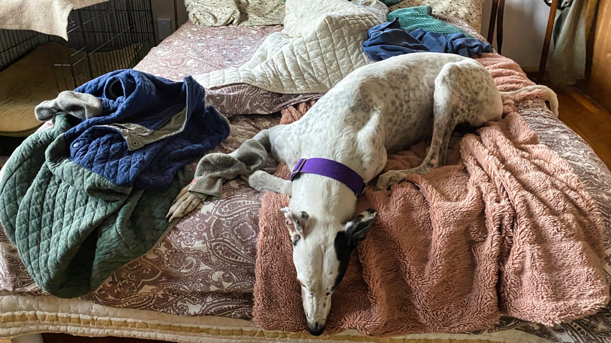
Anyway a sigmoid colon sized piece came out pretty quickly. And I was a bit unsettled that evening but managed a good night’s sleep. Well, Rocky kept one eye open.
